
Can LUMO fNIRS Be Customized for Specific Brain Regions?
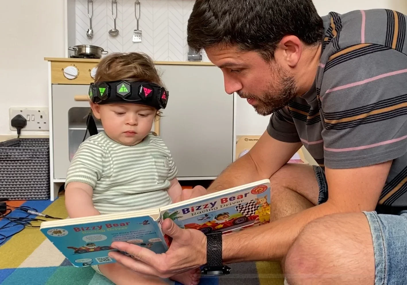
Yes. When needed, LUMO fNIRS can support full-head brain imaging and offer remarkable flexibility in targeting particular cortical regions. Researchers can precisely position sensors over the cortical areas pertinent to their study thanks to its modular HD-DOT architecture, which makes use of interchangeable sensor tiles, custom-built headgear, and configurable docks.
Target Specific Cortical Regions
Researchers can configure layouts for imaging:
Prefrontal cortex
Motor cortex
Visual cortex
Temporal regions
Parietal cortex
Multiple cortical regions simultaneously
This flexibility allows researchers to tailor sensor placement to the specific objectives of each study.
Modular Sensor Configuration
Sensor tiles can be added to dock arrays either completely or partially, enabling anything from thorough whole-head imaging to targeted region-of-interest studies.
Using the same HD-DOT platform, researchers can readily change sensor layouts as their needs change.
Custom Headgear for Different Participant Groups
Custom headgear is available for:
Neonates
Infants
Children
Adults
Headgear can also include bespoke modifications such as cut-outs, adjustable straps, and interchangeable light guides to optimize optical coupling for different experimental requirements and participant populations.
Flexible Brain Imaging for Evolving Research
This level of customization enables researchers to design highly targeted experiments while retaining the flexibility to expand cortical coverage or modify sensor layouts as research objectives change.
Key Customization Features of LUMO fNIRS
Configurable HD-DOT architecture for targeted or whole-head brain imaging.
Precise sensor placement over specific cortical regions.
Supports imaging of the prefrontal, motor, visual, temporal, and parietal cortices.
Configurable dock arrays for region-of-interest or full-head imaging.
Custom headgear available for neonates, infants, children, and adults.
Optional cut-outs, adjustable straps, and interchangeable light guides for optimized optical coupling.
Expand or modify sensor layouts as research objectives evolve without replacing the system.
If you’d like to dive deeper into any particular study or point, feel free to ask us at parag@tidentech.com or tidentech@gmail.com or call us on +91 9987442274.
Tiden Technologies
For both academic and commercial uses, Tiden Technologies is a top supplier of cutting-edge neuroscience, eye tracking, biosensing, and human behavior research technologies.